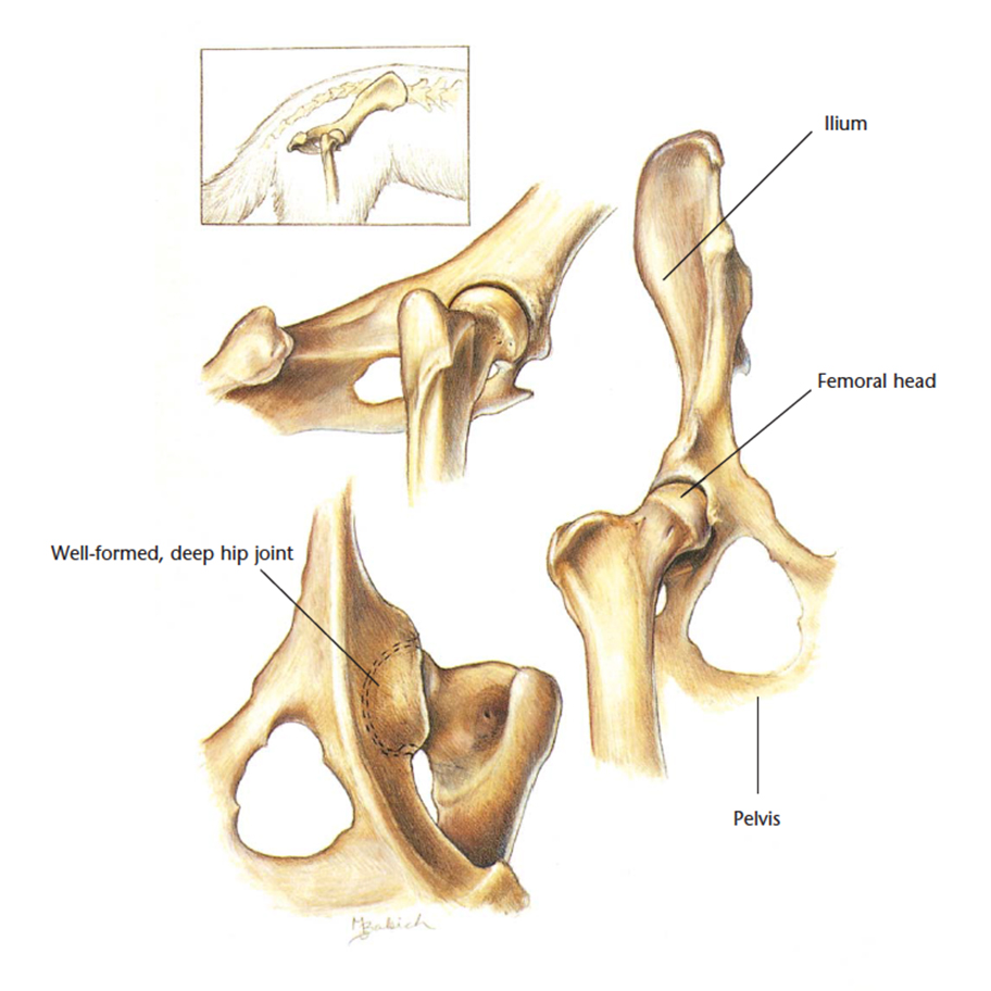
Pelvic Anatomy Dog : Spay and Neuter History and Mystery - MMilani.com : * notice that the kidneys are not labeled on this picture.
byAdmin•
0
Pelvic Anatomy Dog : Spay and Neuter History and Mystery - MMilani.com : * notice that the kidneys are not labeled on this picture.. Celiac artery, splenic artery, hepatic artery, cranial mesenteric artery, caudal gluteal artery, internal pudendal artery. Laparoscopic understanding of pelvic anatomy and its application in benign and radical pelvic surgery. Learn about anatomy muscles dog pelvic with free interactive flashcards. Dog anatomy a pictorial approach to canine structure. There are many organs that sit in the pelvis, including much of the urinary system, and lots of the male or female reproductive systems.
Lotze, md facog female pelvic medicine & reconstructive surgery division & fellowship director, women's pelvic health & continence center clinical. Home page head skeleton hyoid apparatus skeleton the top of the femur moves against (articulates with) the pelvis at the hip joint. The detailing of these structures changes based on dog breed due to the huge variation of size in dog. 12 photos of the pelvic bone anatomy. Branches of the internal iliac artery.
Hills Pet Nutrition | VetCheck Cat Pelvis Anatomical Diagram from vetcheck.it Veterinary, anatomy, dog, muscles, thoracic limb, (1 of 3). Dog anatomy comprises the anatomical studies of the visible parts of the body of a domestic dog. Pelvic floor anatomy & function: The poster shows the superficial muscles, skeletal system with surface anatomy. Pictured above shows the dog muscle anatomy of the canine. Some canine anatomical names may be familiar to you — dogs have elbows and ears and eyes — but many anatomical terms used to describe parts of a dog are similar to the ones used for horses. Anatomy of the pelvic floor. There are many organs that sit in the pelvis, including much of the urinary system, and lots of the male or female reproductive systems.
Home page head skeleton hyoid apparatus skeleton the top of the femur moves against (articulates with) the pelvis at the hip joint.
Details of structures vary tremendously from breed to breed, more than in any other animal species, wild or domesticated, as dogs are highly variable in height and weight. 100 x 100 png 10 кб. The detailing of these structures changes based on dog breed due to the huge variation of size in dog. Veterinary, anatomy, dog, muscles, thoracic limb, (1 of 3). Learn about the blood vessels, organs, nerves and peritoneal cavity. This particular dog uses bully max™ when it comes to the pelvic limb region of the body, there are another seven muscles/muscle groups. 3d interactive models and tutorials on the anatomy of the abdomen and pelvis. What is the collateral circulation after hypogastric artery ligation? Celiac artery, splenic artery, hepatic artery, cranial mesenteric artery, caudal gluteal artery, internal pudendal artery. Blood supply of the male pelvis. The poster shows the superficial muscles, skeletal system with surface anatomy. 216 x 200 jpeg 9 кб. Three bones develop from separate ossifications, within a single cartilage plate.
Mri studies have outlined the anatomy of pelvic floor muscles much more clearly than was possible with anatomic. The pelvic girdle consists of two symmetrical halves. Branches of the internal iliac artery. 3d interactive models and tutorials on the anatomy of the abdomen and pelvis. Pictured above shows the dog muscle anatomy of the canine.
Dog Skeletal Anatomy Poster from www.acupunctureproducts.com Mri studies have outlined the anatomy of pelvic floor muscles much more clearly than was possible with anatomic. Details of structures vary tremendously from breed to breed, more than in any other animal species, wild or domesticated, as dogs are highly variable in height and weight. Surgical pelvic anatomy in gynecologic oncology. Veterinary, anatomy, dog, muscles, thoracic limb, (1 of 3). There are many organs that sit in the pelvis, including much of the urinary system, and lots of the male or female reproductive systems. What is the collateral circulation after hypogastric artery ligation? Dog anatomy poster created using vintage images. Pelvic anatomy mri variant anatomy pelvic viscera.
Branches of the internal iliac artery.
How tho find the pssoas minor, pssoas major, qudratus lumburum and ilioc musceles. Canine pelvic limb anatomy by *leonca on deviantart | dog. The kidneys are tucked up close to the liver toward the spine. Mri studies have outlined the anatomy of pelvic floor muscles much more clearly than was possible with anatomic. Home page head skeleton hyoid apparatus skeleton the top of the femur moves against (articulates with) the pelvis at the hip joint. Pictured above shows the dog muscle anatomy of the canine. Muscle, organ and skeletal anatomy). There are many organs that sit in the pelvis, including much of the urinary system, and lots of the male or female reproductive systems. This is pelvic anatomy laparoscopic hysterectomy by irocket on vimeo, the home for high quality videos and the people who love them. Dog anatomy details the various structures of canines (e.g. The pelvic girdle consists of two symmetrical halves. * notice that the kidneys are not labeled on this picture. 3d interactive models and tutorials on the anatomy of the abdomen and pelvis.
In this video we are talking about the anatomy of the pelvis of the horse and the main differences between the main domestic. Cardiovascular system of the cat. Branches of the internal iliac artery. 216 x 200 jpeg 9 кб. Blood supply of the male pelvis.
Skeleton of the cat | Pelvic girdle, Hip bones, Os coxae from i.pinimg.com How tho find the pssoas minor, pssoas major, qudratus lumburum and ilioc musceles. 12 photos of the pelvic bone anatomy. The pelvic girdle consists of two symmetrical halves. Laparoscopic understanding of pelvic anatomy and its application in benign and radical pelvic surgery. Veterinary, anatomy, dog, muscles, thoracic limb, (1 of 3). Muscle, organ and skeletal anatomy). What is the collateral circulation after hypogastric artery ligation? Canine pelvic limb anatomy by *leonca on deviantart | dog.
Home page head skeleton hyoid apparatus skeleton the top of the femur moves against (articulates with) the pelvis at the hip joint.
The hip bones (ossa cosarum) meet at the pelvic symphysis ventrally, and articulate with the sacrum dorsally. Dog anatomy poster created using vintage images. Learn about the blood vessels, organs, nerves and peritoneal cavity. Branches of the internal iliac artery. Lotze, md facog female pelvic medicine & reconstructive surgery division & fellowship director, women's pelvic health & continence center clinical. Show the clinical relevance of anatomy in such a way is a powerful tool for stimulating students' interest. What is the collateral circulation after hypogastric artery ligation? * notice that the kidneys are not labeled on this picture. Pelvic floor anatomy & function: Laparoscopic understanding of pelvic anatomy and its application in benign and radical pelvic surgery. Surgical pelvic anatomy in gynecologic oncology. This particular dog uses bully max™ when it comes to the pelvic limb region of the body, there are another seven muscles/muscle groups. Some canine anatomical names may be familiar to you — dogs have elbows and ears and eyes — but many anatomical terms used to describe parts of a dog are similar to the ones used for horses.
Agreements & disagreements workshop 36 pelvic anatomy. This is pelvic anatomy laparoscopic hysterectomy by irocket on vimeo, the home for high quality videos and the people who love them.